An echocardiogram uses sound waves to produce images of your heart. This common test allows your doctor to see your heart beating, pumping blood and heart valvular condition. Your doctor can use the images from an echocardiogram to identify heart disease. Echocardiography could identify changes in your heart size, weakened or damaged heart valves, high blood pressure or other diseases can cause the chambers of your heart to enlarge or the walls of your heart to be abnormally thickened, heart defects. An echocardiogram can show problems with the heart chambers, abnormal connections between the heart and major blood vessels, and complex heart defects that are present at birth.
 |
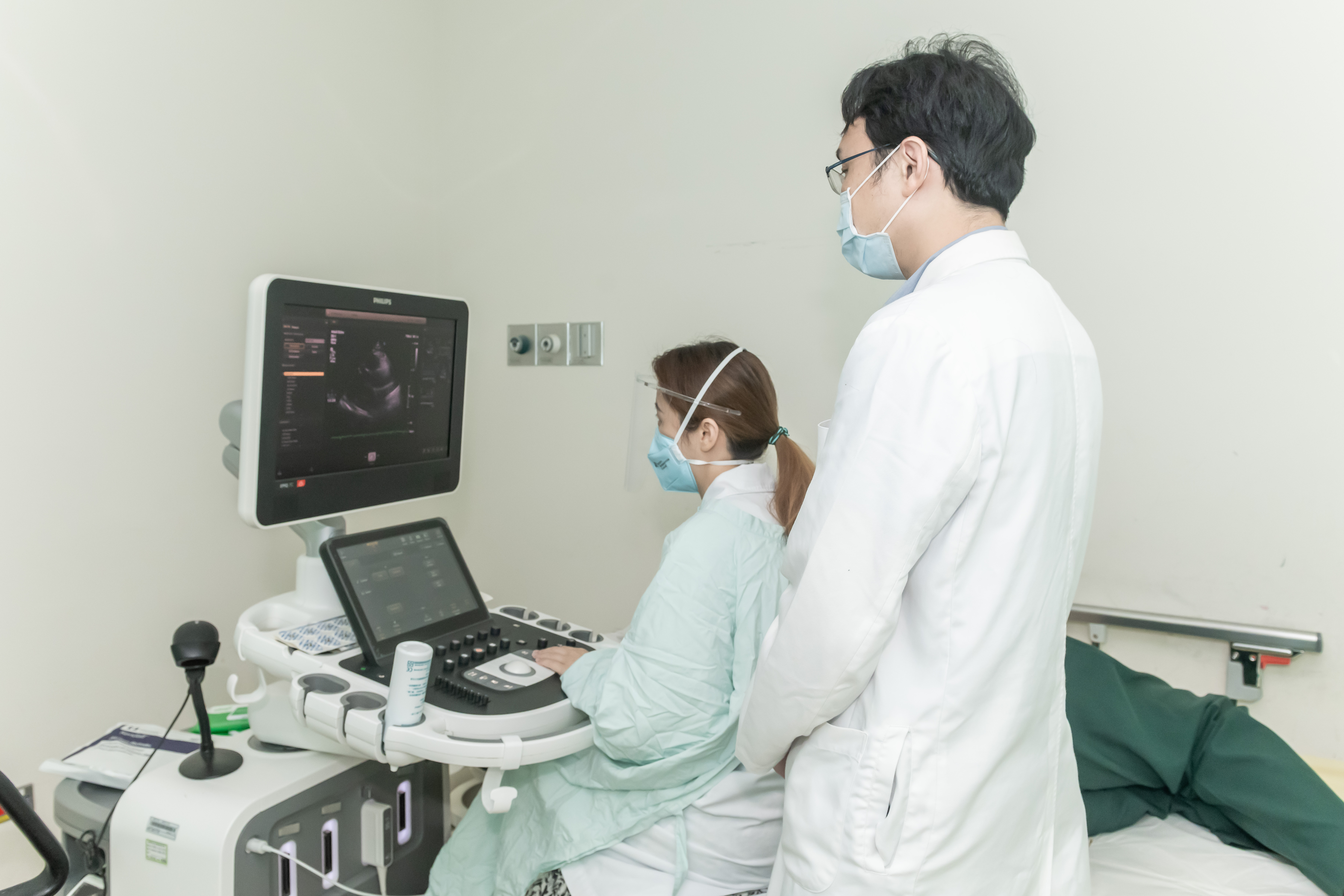 |
Transthoracic echocardiogram
In this standard type of echocardiogram:
A technician (sonographer or doctor) spreads gel on a device (transducer).
The sonographer presses the transducer firmly against your skin, aiming an ultrasound beam through your chest to your heart.
The transducer records the sound wave echoes from your heart.
A computer converts the echoes into moving images on a monitor.
Transesophageal echocardiogram
If your doctor wants more-detailed images or it's difficult to get a clear picture of your heart with a standard echocardiogram, your doctor may recommend a transesophageal echocardiogram.
In this procedure:
Your throat will be numbed.
A flexible tube containing a transducer is guided down your throat and into your stomach (esophagus). The transducer records the sound wave echoes from your heart. A computer converts the echoes into detailed moving images of your heart, which your doctor can view on a monitor.